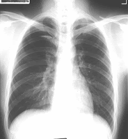
Chest X-Ray
What is Chest X-ray
The chest x-ray is the most commonly performed diagnostic x-ray examination. A chest x-ray makes images of the heart, lungs, airway, blood vessels and the bones of the spine and chest. During an x-ray, a focused beam of radiation is passed through your body, and a black-and-white image is recorded on special film or a computer. The x-ray image that is created looks like a negative from a black and white photograph.
X-rays work because the body's tissues vary in density (thickness).
Each tissue allows a different amount of radiation to pass through and expose the x-ray sensitive film, which results in a shadow image of the organ. Bones, for example, are very dense, and most of the radiation is prevented from passing through to the film. As a result, bones appear white on an x-ray. Tissues that are less dense--such as the lungs, which are filled with air--allow more of the x-rays to pass through to the film and appear on the image in shades of gray.
A chest x-ray can be used to define abnormalities of the lungs such as excessive fluid, pneumonia, bronchitis, asthma, cysts, and cancers. Heart abnormalities, including fluid around the heart (pericardial effusion), an enlarged heart, heart failure, or abnormal anatomy of the heart can be revealed on the films.


1 comment:
Provide very meaningful information about Chest x-ray. Thanks for sharing.
formation rayons-x sécurité
Thanks.
Post a Comment