Chest X-Ray


What is Chest X-ray
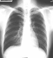
The chest x-ray is the most commonly performed diagnostic x-ray examination. A chest x-ray makes images of the heart, lungs, airway, blood vessels and the bones of the spine and chest. During an x-ray, a focused beam of radiation is passed through your body, and a black-and-white image is recorded on special film or a computer. The x-ray image that is created looks like a negative from a black and white photograph.
X-rays work because the body's tissues vary in density (thickness).

Each tissue allows a different amount of radiation to pass through and expose the x-ray sensitive film, which results in a shadow image of the organ. Bones, for example, are very dense, and most of the radiation is prevented from passing through to the film. As a result, bones appear white on an x-ray. Tissues that are less dense--such as the lungs, which are filled with air--allow more of the x-rays to pass through to the film and appear on the image in shades of gray.
A chest x-ray can be used to define abnormalities of the lungs such as excessive fluid, pneumonia, bronchitis, asthma, cysts, and cancers. Heart abnormalities, including fluid around the heart (pericardial effusion), an enlarged heart, heart failure, or abnormal anatomy of the heart can be revealed on the films.
Readmore ..»»

What are some common uses of the procedure ?
The chest x-ray is performed to evaluate the lungs, heart and chest wall. A chest x-ray is typically the first imaging test used to help diagnose symptoms such as:
• Lung disorders such as pneumonia, emphysema, tuberculosis and lung cancer
• Heart disorders such as congestive heart failure (which causes the heart to enlarge)
• Fractures (breaks) of the bones in the chest, including the ribs and collarbone, as well as breaks in the bones of the upper spine
• Reasons for shortness of breath, a bad or persistent cough, or chest pain

Who performs the test?
A radiology technologist, a skilled medical professional who is trained in x-ray procedures, will perform the test. A radiologist, a doctor who specializes in evaluating x-rays and other radiology procedures, will interpret the x-rays and report the test results to the doctor.

Readmore ..»»

What are the risks of a chest x-ray ?
Chest x-rays expose the patient briefly to a minimum amount of radiation. Any radiation exposure has some risk to the tissues of the body. The radiation exposure in a chest x-ray is minimized by the type of x-ray high speed film, which does not require as much radiation exposure as in the past.
Women who are pregnant, especially in early pregnancy, should notify their physicians, as the fetus is at risk for harm with any radiology technique.

How do I prepare for the test ?
There is no special preparation for a chest x-ray. It is important to tell the technologist if you are or may be pregnant. X-rays generally are not used on pregnant women because of the possible risk of radiation exposure to the developing baby.
Before the test begins, you will be asked to remove your clothing usually just from the waist up and put on a hospital gown. You also will be asked to remove all jewelry and any other objects containing metal (such as eyeglasses and hair pins). This is done because metal can block the image and interfere with the test results.

Readmore ..»»

What are the benefits versus risks?
Benefits
  • No radiation remains in a patient's body after an x-ray examination.
  • X-rays usually have no side effects.
  • X-ray equipment is relatively inexpensive and widely available in physician offices, ambulatory care centers, nursing homes and other locations, making it convenient for both patients and physicians.
  • Because x-ray imaging is fast and easy, it is particularly useful in emergency diagnosis and treatment.
Risks
  • There is always a slight chance of cancer from radiation. However, the benefit of an accurate diagnosis far outweighs the risk.
  • The chest x-ray is one of the lowest radiation exposure medical examinations performed today. The effective radiation dose from this procedure is about 0.1 mSv, which is about the same as the average person receives from background radiation in 10 days. See the Safety page for more information about radiation dose.
  • Women should always inform their physician or x-ray technologist if there is any possibility that they are pregnant. See the Safety page for more information about pregnancy and x-rays.
What are the limitations of Chest X-ray?
The chest x-ray is a very useful examination, but it has limitations. Because some conditions of the chest cannot be detected on an x-ray image, this examination cannot necessarily rule out all problems in the chest. For example, very small cancers may not show up on a chest x-ray. A blood clot in the lungs, a condition called a pulmonary embolism, cannot be seen on chest x-rays.


Readmore ..»»